

 ع
ع


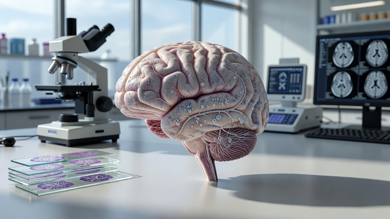
The Sudha Gopalakrishnan Brain Centre at the Indian Institute of Technology Madras has produced what it describes as the world’s most detailed three-dimensional atlas of the human brainstem at cellular resolution, according to a BBC News report published on July 13, 2026. Named Anchor, the atlas combines more than 500 tissue sections from foetal, childhood and adult brains and identifies over 200 clusters of brain cells along with associated nerve pathways. It relies on high-resolution microscope images and eight chemical markers rather than more expensive molecular methods, creating a digital resource that allows users to navigate from whole-brain MRI scans down to individual neurons while preserving spatial relationships. The centre made the atlas freely available online to serve as a reference for researchers, neurologists and neurosurgeons around the world.
This brainstem map addresses a longstanding gap in neuroscience because the region, though only a small portion of the brain, controls essential functions such as breathing, heartbeat, sleep and movement, the BBC News report stated. Damage to specific cell clusters there can lead to catastrophic outcomes, yet its dense structure has made high-resolution mapping difficult for decades. The project involved around 20 scientists who spent 18 months analysing more than 200 brain sections to integrate MRI data, microscopic anatomy and three-dimensional reconstruction. Mohanasankar Sivaprakasam, who heads the centre, told the BBC News that detailed studies of human brain tissue remain scarce even though animal brains have been mapped extensively.
Rebecca Folkerth, affiliated with Harvard Medical School and New York University and a collaborator on the project, described the atlas as fulfilling a long-held ambition in neuropathology, the BBC News report noted. “What the Indian centre has created is essentially what I dreamed of early in my career – to have brain scans match the brain’s microscopic anatomy,” Folkerth, who has examined thousands of brains over more than three decades, told the BBC. The atlas is not a diagnostic tool but could help compare healthy and diseased tissue to improve understanding of conditions including Parkinson’s disease, stroke, Alzheimer’s disease and sudden infant death syndrome. Partha Mitra of Cold Spring Harbor Laboratory, who has also worked with the centre, said such detailed atlases could have a transformative impact on neurological disease research by revealing cell-by-cell differences.
This Indian initiative takes place amid a series of global brain-mapping projects that have accelerated in recent years, a review by the Human Brain Project indicated. The European effort, which ran from 2013 to 2023, produced digital brain atlases, modelling tools and simulation platforms now accessible through the EBRAINS infrastructure, according to project documentation. In the United States the BRAIN Initiative Cell Atlas Network released its first major human brain data sets in October 2024, building on earlier mappings of mouse and primate brains, the Allen Institute for Brain Science reported. These international activities underscore how progress in the field now depends on engineering, computation and large-scale collaboration as much as traditional biology.
India faces a substantial burden from neurological disorders that could benefit from such mapping resources, according to World Health Organization figures. The organisation reported that 57 million people lived with dementia worldwide in 2021, with more than 60 percent of cases in low- and middle-income countries and nearly 10 million new cases emerging each year. A study published in the journal Alzheimer’s & Dementia placed dementia prevalence among Indian adults aged 60 and older at 7.4 percent, equating to an estimated 8.8 million affected individuals, with higher rates observed in rural areas and among those with lower education levels. The Sudha Gopalakrishnan Brain Centre now plans to image more than 100 whole human brains across life stages and disease states including Alzheimer’s and dementia, expanding on its earlier completion of second-trimester foetal brain maps, a November 2025 announcement from the centre detailed.
The project originated in 2017 with encouragement from IT entrepreneur Kris Gopalakrishnan and moved into formal development in 2020 after the centre created its own advanced imaging technology, according to statements from Sivaprakasam at an Indian Academy of Neurosciences meeting. “The brain is extraordinarily complex, comprising over 100 billion cells and trillions of connections,” Sivaprakasam said in remarks carried by The Indian Practitioner. “Today, magnetic resonance imaging allows us to visualize the brain only at a millimeter level. Our work aims to capture the human brain at the resolution of every single cell.” Shubha Tole of the Tata Institute of Fundamental Research called the programme a visionary effort that places India at the international table through its integration of engineering, neuroscience and medicine, the BBC News report added.